

vol.
IV08/12 y.
issue 2
Endobronchial ultrasonography – a new possibility in interventional pulmology
Abstract
Diagnosis of the nature of the peripheral pulmonary and mediastinal lesions as well as exact staging the non-small cell lung cancer are a significant challenge for pulmonologists. The possibilities of fibrobronchoscopy are limited because of the fact that information is obtained about endobronchial alterations, and an indirect one only about changes in the bronchial wall and outside. In spite of its high information value, computer tomography and positron emission tomography can prove to be misleading when defining the nature of the lesions in the pulmonary parenchyma and mediastinum as well. Endobronchial ultrasonography (EBUS) with or without transbronchial needle biopsy is a new diagnostic method enlarging the possibilities for evaluation of the alterations in the bronchial wall and neighbour structures and warranting a morphological material from the pathologically changed mediastinal lymph nodes, especially when staging the non-small cell lung cancer without resorting to the ‘gold standard, i.e. to mediastinoscopy or mediastinotomy as well as when defining the nature of the peripheral lesions. Linear EBUS provides real-time biopsies of centrally located pulmonary and mediastinal processes while radial one reveals the depth of bronchial wall involvement by the tumour process and helps the diagnosis of the peripheral lesions and interventional therapeutic endobronchial procedures. The method is minimally invasive, of good tolerability on the patient’s part and minimal complications in comparison with the other invasive diagnostic procedures.
1. Clinic of Pulmology and Phthisiatry, St. Marina University Hospital of Varna
Diagnosis of the mediastinal formations, mediastinal lymphadenomegaly and peripheral pulmonary lesion still represents a significant challenge for pulmonologists and chest surgeons. Morphological diagnosis is related to the wide application of the fine needle aspiration biopsy, transthoracic aspiration biopsy, mediastinoscopy that is the ‘gold standard’ in the diagnosis of the mediastinal pathology, and video-assisted thoracoscopy. These methods present with their advantages and disadvantages concerning the obtained tissue material, safety and price. Bronchoscopy plays an important role in lung cancer diagnosis and staging. Endobronchial biopsy under visual control enables an exact diagnosis in more than 90% of the cases, however, in peripheral tumors without any visual access things are not the same. Introduction of computer tomography (CT) considerably improved the possibilities for lung cancer diagnosis and staging; however, the experience gained concerning its reliability when determining the mediastinal involvement and the degree of bronchial wall infiltration proved to be disappointing. In approximately 100% of the patients with non-small cell lung cancer staged by means of CT, explorative thoracotomy ends without any resection because of an advanced process remaining non-detected preoperatively. Besides 25-35% of the patients with a resection appearing successful present with an early postoperative relapse (32). Introduction of positron- emission tomography (PET-CT) improved the possibilities for staging, however, without the necessary categoricalness.
It has been established that not only tumor- involved but also inflammatorily changed lymph nodes can present with an increased extent of metabolism as those of a normal size and metabolism can contain tumor cells (10). Konishi et al. (26) compare the role of CT and PET-CT when staging the non-small cell lung cancer and establish a sensitivity of 55% and specificity of 96% in CT towards 73% and 98% in PET-CT, respectively. Falsely positive results of PET-CT are related to other pulmonary diseases such as interstitial pneumonitis, tuberculosis, silicosis, etc.
Fig. 1. “Peripheral”and “central” EBUS
a) “Peripheral” EBUS – tiny probe UM-S20-20R without balloon sleeve on top for the diagnosis of peripheral lung lesions.
b) “Central” EBUS – balloon probe UM-BS20-26R with balloon in the distal part, allowing circular contact of the ultrasound and image from 360 degrees of peritracheal and peribronchial structures.

Endoscopic ultrasound (EUS) starts its development in the 80-ies of the 20th century as an integral part of the examination of the gastrointestinal tract by using of a radial probe. Esophagus’ anatomical location behind and to the left of trachea defines EUS application when staging the lung cancer as well as for the performance of fine- needle aspiration biopsy (FNAB) of lymph nodes and adrenal gland. In 1993, Wiersema et al. (33) published the first EUS application for staging the non-small cell lung cancer by means of FNAP through the esophagus. The complete mediastinal visualization by EUS is restricted because of airway interference, on the one hand, and of the difficult lymph node accessibility in the positions of 2Р, 3 and 4Р, on the other. These circumstances are the precondition for the development of the idea of the ultrasonography of the airways.
The history of endobronchial ultrasonography (EBUS) begins with the communication by Hurter and Hanrath (22) about EBUS of the lung and mediastinum in 1990. The development and expansion of EBUS application continues in Germany, Japan, and the USA as one makes use of endovascular microprobes (7, 13, 22). Becker (8) elaborates a flexible catheter with an ultrasonic probe for insertion into the large airways by means of a balloon on its tip with a view of ensuring a circular contact with the bronchial wall. Nowadays, two models of EBUS, i.e. radial and linear, are applied for different purposes.
Radial EBUS
Radial EBUS is provided with a rotation transducer of 20 MHz with possibility of 12-30 MHz that can be inserted with or without guide sheath through the working canal of a standard fibro- bronchoscope of 2-2.8 mm. The radial probe is of different size and of an external diameter of 1.4-2.6 mm.
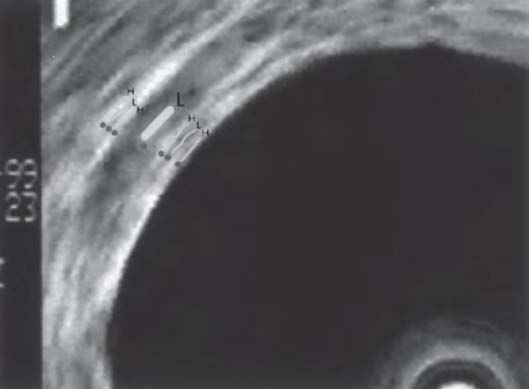
Fig. 2. Layers of the bronchial wall visualized by 20 MHz probe (enlarged image).
From the inside out are: mucosa (H), submucosa (L), endochongrium (H), cartilage (L), perichondrium (H), connective tissue (L), and adventitsiya. L – low-density layer, H – high-density layer.
The ‘central’ probe, Balloon Probe UM-BS20-26R is with a polyethylene sheath and latex balloon in its distal part. The contact between the bronchial wall and transducer is accomplished though filling-up of the latex balloon with physiological serum enabling a better contact in the proximal airways with a view to inspect the bronchial wall or to perform a transbronchial biopsy (TBB) of the lymph nodes.
The ‘peripheral’ EBUS (Miniature Probe UM-S20- 20R) is without any balloon sheath and is used for identification of the peripheral pulmonary lesions with the purpose of a biopsy (Fig. 1).
It is not necessary to stimulate the contact with the bronchial wall within the distal airways because they ensure a sufficient contact between the transducer and bronchial mucosa. With the radial EBUS there is an image of 360o that is perpendicular to the long axis of the airways. Image resolution is of 1 mm but depth of access is of 4-5 cm.
Fig. 3. Findings in СТ and EBUS:
On the left above – CT finding of central infiltrative formation.
On the right above – EBUS – intact bronchial wall. Below – enlarged EBUS image.
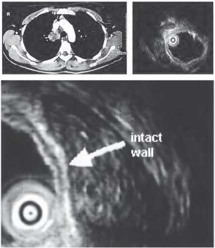
Fig. 4. Histological and EBUS findings:
On the left above – EBUS – tumor infiltration of tracheal wall.
On the right above – corresponding histological material – destruction of the cartilage.
Below – enlarged EBUS image – tumor infiltration of the bronchial wall (left main bronchus).
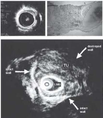
Usually, radial EBUS is carried out following a standard fibrobronchoscopy (FBS) with inspection of the bronchial tree. After finding the image, both echoscopic and endoscopic images are compared by means of reference points such as large vessels (anechogenic), vertebral column, and esophagus. Tumor formation looks hypoecho- genic when compared to the healthy lung tissue and there are clearly outlined borderlines between the lesion and the aerated lung. After identification of the target lesion, the probe moves to it and TBB is undertaken. At the same time, the biopsy guided by the radial EBUS is restricted because of the absent real-time image. The diagnostic data described about TBB with the radial EBUS vary between 72% and 86% as this procedure demonstrates an improvement of the diagnostic level over the blind conventional TBB concerning the mediastinal lymph nodes at positions of 2, 3, and 4 (14). The radial EBUS enables the examination of any bronchial wall layers, more than 7 ones (Fig. 2). They are the following: mucosa, endochondrium, perichondrium and adventitia, which are hyperechogenic, and submucosa, cartilaginous tissue and connective tissue, which are hypoechogenic (1, 2). EBUS displays better than CT the extent of infiltration or compression by the tumour presenting with a sensitivity of 89% and specificity of 100% (Figs. 3 and 4). The patient can be out of range of the invasive therapeutic procedure such as photodynamic therapy or brachy- therapy through identification of evidence about invasion in the cartilaginous tissue of the tumor in favor of the surgical intervention while through visualization of a nearly located blood vessel prior to debridement or thermal abrasion can avoid fatal bleeding.
Indications for radial EBUS application
Early central lung cancer
With small roentgenologically invisible tumors, the decision for a local endobronchial therapeutic intervention depends on their intraluminal and intramural scope of the various bronchial wall layers (2). In contrast to the x-ray examination, by means of EBUS one can analyze very small-sized tumors and differentiate them from benign neoplasms. Herth et al. (21) demonstrate how EBUS improves the specificity that forecasts malignancy by 50% up to 90% in autofluorescence-positive lesions which are invisible when standard FBS with white light is applied. The combination of EBUS and autofluorescence can be an effective approach for endobronchial treatment of malignant neoplasms limited within the bronchial wall (15, 16, 28). EBUS differentiates the tumour invasion from the compression of the tracheobronchial wall in 94% while CT does in 51% of the cases. During the preoperative staging EBUS is used for determination of the resection margin as it warrants a detailed analysis of the intraluminal, submucous and intramural tumor dissemination (17).
Advanced lung cancer
Several authors accept that the radial EBUS can be used as a means in the interventional bronchology that contributes to the restoration of the pass ability of the large airways through laser coagulation or stenting as well as to the determination of the status of the vessels in the proximity of the tumors prior to their destruction (15, 30). Within a large trial, a total of 2 446 patients have undergone a therapeutic bronchoscopy as in 1 174 of them it has been done by means of a radial EBUS. In 43% of the cases it is capable of modifying the therapeutic procedures by adapting the stent size, of finalizing the tumor debridement closely to the vessels and thus of more exactly performing the endoscopic treatment. In the cases of laser treatment, EBUS application restricts the bleeding from the nearly located vessels (6).
Diagnosis of the peripheral lesions
Malignant peripheral lesions are, usually, hy- poechogenic and of well-defined peripheral outlines while an air bronchogram is absent (15, 27) (Fig. 5).
The histological diagnosis of the peripheral pulmonary lesions by using of FSB under fluoroscopic control, or the biopsy under CT-control is a routine procedure. Herth et al. (15) prove that a similar procedure under EBUS-control presents with the same success and a reliability level of ap- proximately75% provided that there are considerably less complications. Kurimoto et al. (29) apply a radial EBUS in 150 patients presenting with a solid peripheral nodule. A specific diagnosis is achieved in 116 patients (in 77% of the cases). The results are even better (in 87% of the cases) if the probe is situated in the lesion itself rather than in its proximity.
Fig. 5. Lung nodule.
(А) СТ – Lung nodule (18 mm) in the right middle lobe.
(В) EBUS – Hypoechogenic nodule with hyperechogenic areas.
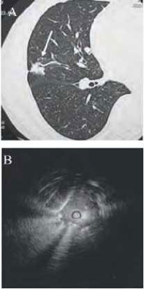
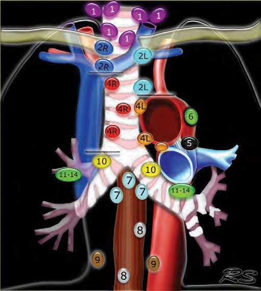
Fig. 6. Map of regional lymph nodes in staging lung cancer, adapting according American Thoracic Society.
In Paone’s et al. trial (31), 206 out of a total 293 patients have been randomized either for a routine FSB, or for radial EBUS. Positive results have been obtained in 76% of the patients who have undergone EBUS procedure and in 52% of the cases following FSB under fluoroscopic control. When the peripheral lesion is larger than 3 cm there are no differences while when it is smaller than 3 cm the sensitivity and accuracy of EBUS are of 75% and 83%, respectively, and of 31% and 50% after the routine examination. The diagnostic reliability of TBB guided during EBUS concerning the peripheral pulmonary lesions sized below 20-30 mm is of 61-80% (Table 1).
Lymph node staging
Non-small cell lung cancer staging is of significant importance when selecting the therapeutic approach. By means of EBUS, 2-3 mm-sized lymph nodes can be established and their structure can be defined. The results from the trans- bronchial needle biopsy (TBNA) can be significantly improved through the endosonographic lymph node localization – up to more than 80%, especially in the cases of upper and lower para- tracheal lymph nodes that are inaccessible by using of CT. Herth et al. (18) compare the results from the TBNA under EBUS-control and from the conventional biopsy, which are of diagnostic information value in 85% and in 66% of the cases. The additional analysis of lymph node positions reveals that in case of localization outside the endoscopic range, i. e. positions of 2, 3, and 4, EBUS enhances the number of the positive results (Fig. 6). On the other hand, with enlarged subcarinal lymph nodes, both the conventional biopsy and TBNA guided during EBUS show similar results.
Table 1. Radial EBUS when performing TBB from peripheral pulmonary lesions (< 20-30 mm) with a diagnostic accuracy of 61 -80% (3).
| Authors | Technique | Number of lesions | Mean size of lesions (mm) | Diagnostic reliability (%) |
| Paone et al. (32) | EBUS-TBB | 25 | < 20 | 71 |
| 47 | < 30 | 75 | ||
| 40 | > 30 | 83 | ||
| Yamada et al. (35) | EBUS-TBB by guide sheath | 158 | 21 ± 6 | 67 |
| 40 | < 15 | 40 | ||
| 118 | > 15 | 76 | ||
| Yoshikava et al. (36) | EBUS-TBB and brush biopsy by guide sheath (without fluoroscopy) | 123 | 31 ± 16 | 62 |
| 37 | < 20 | 30 | ||
| 86 | > 20 | 76 | ||
| Chao et al. (9) | EBUS-TBNA, clip biopsy and BL | 88 | 35 ± 10 | 78 |
| EBUS-clip biopsy and BL | 94 | 35 ± 8 | 61 | |
| Kikuchi etal. (25) | EBUS – by guide sheath and fluoroscopy±curettage-TBB/brush biopsy | 24 | < 30 | 58 |
| 15 | < 20 | 53 | ||
| 9 | 20-30 | 67 | ||
| Asahina etal. (4) | EBUS by guide sheath, navigation by virtual bronchoscopy, fluoroscopy ±curettage-TBB/brush biopsy | 30 | < 30 | 63 |
| 18 | < 20 | 44 | ||
| 12 | 20-30 | 92 | ||
| Kurimoto etal. (30) | EBUS – by guide sheath and fluoroscopy±curettage-TBB/brush biopsy | 81 | 20 | 73 |
| Asano et al. (5) | EBUS-TBB and brush biopsy by guide sheath | 32 | 21 | 84 |
| Koh et al. (26) | EBUS-TBB by guide sheath | 29 | 35 | 62 |
| Fielding etal. (12) | EBUS-TBB by guide sheath | 140 | 29 | 66 |
| Dooms etal. (11) | EBUS-TBB | 50 | 37 ± 20 | 84 |
Linear EBUS-TBNA (Convex)
It represents a bronchoscope with an integrated curvilinear electron transducer on the tip that enables the real-time TBNA performance (Fig. 7). Its outer diameter is of 6.9 mm but the inner one is of 2 mm which makes it of larger volume than the common fibrobronchoscope and requires an oral manner of introduction. The angle of inspection is of 80o and the direction of the image is of 35o. Ultrasound (US) frequency of 7.5 MHz ensures a penetration into a depth of up to 5 cm. The transbronchial needle of 22 Gauge is inserted into the working canal of the fibrobronchoscope.
US-image is obtained either through probe placement directly to the trachea or bronchial wall, or through instillation of physiological serum in a balloon at the tip of the probe, which can improve image quality. US-image can be kept that enables the two-dimensional examination of the lesion. It is comfortable that both US- and bronchoscopic light images can simultaneously be observed. An additional Doppler examination is capable of differentiating the tissues from vascular structures and, in this way, of avoiding the puncture of a vessel. The relatively wide internal canal of the needle enables the aspiration of a material suitable for cytological and histological examination as the recommended number of the reiterated procedures is 3. The linear BUS is recommended predominantly for the examination of the mediastinal lymph nodes at the positions of the upper paratracheal ones – 1, 2R and 2L, lower paratracheal ones – 4 and 4L, subcarinal ones – 7, hillar ones – 10 as well as interlobar ones – 11, for the diagnosis of the mediastinal and pulmonary tumors. The paraaortic (6), aortic-pulmonary, subaortic (5), paraesophageal (8) and pulmonary ligament lymph nodes (9) are inaccessible for this technique.
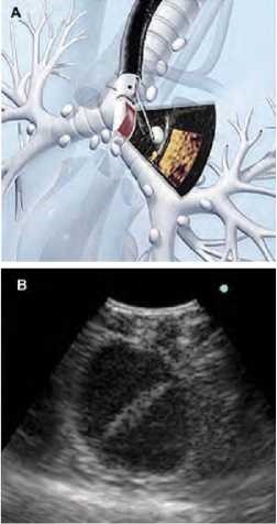
A basic indication for real-time EBUS-TBNA is the examination of the enlarged mediastinal lymph nodes (1) as established after CT of the chest aiming at staging the non-small cell lung cancer (Fig. 8).
Herth et al. (19) diagnose 534 out of a total of 572 punctured lymph nodes (94% of the cases). Biopsies have been carried out from all the areas rich in lymph nodes such as 2L, 2R, 3, 4R, 4L, 7, 10R,11R, and 11L of a mean size of 1,6 cm. The established sensitivity is of 94% and specificity is of 100%. The method is useful in the cases with CT- not enlarged lymph nodes, too. By using of EBUS- TBNA, Herth et al. (20) examine 100 lung cancer patients without any enlarged mediastinal lymph nodes and juxtapose the results with its surgical proof. The sensitivity of EBUS-TBNA is of 92%, the specificity is of 100% and the negative predicting value is of 96%.
EBUS-TBNA can successfully be applied in adenopathies of other etiology. A diagnostic sensitivity of 91% is achieved when proving the clinically suspicious lymphoma (23). The method displays a sensitivity of 85-94% concerning the primary diagnosis of sarcoidosis. TBB being most commonly used demonstrates positive results in 46-90% as in 5% of the cases it is accompanied by bleeding or pneumothorax (3).
Fig. 7. Convex probe EBUS.
(A) The tip of the convex probe (Olympus XBF-UC180-DT8) has a curved linear transducer of 7.5 MHz.
(B) Balloon attached on the top of bronchoscope is filled with saline.
(C) Transbronchial aspiration needle is inserted through the working channel.
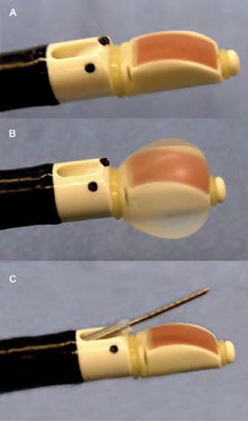
Fig. 8. Convex probe EBUS.
(A) Convex probe EBUS in a main bronchus.
(B) Ultrasound image – biopsy needle in a lymph node.
Conclusion
During the recent several years, EBUS has established itself as a precise and reliable diagnostic method. Numerous investigations and scientific communications confirm its high diagnostic value with a series of pulmonary and mediastinal diseases as well as with staging the non-small cell lung cancer. Its different variants possess their advantages: the linear EBUS ensures real-time biopsies in centrally located pulmonary and mediastinal processes while the radial one reveals the depth of bronchial wall involvement by the tumor process and helps the diagnosis of the peripheral lesions and the interventional therapeutic endobronchial procedures. The method is minimally invasive and of a good tolerability on the part of the patient as well as related with minimal complications when compared to the other invasive diagnostic procedures.
References
- Петрова, Д., О. Георгиев, Ц. Мондешки, Д. Вълев. Възможности и значение на ендобронхиалната ултразвукова диагностика. Пневмол. и фтизиатр., 40, 2005, № 1, 51-55.
- Петрова, Д., О. Георгиев, Р. Билюков, Ц. Мондешки. Съвременни диагностични техники в бронхоскопията за ранна диагноза на белодробния карцином. InSpiro, 3, 2010, № 3, 6-11.
- Anantham, D., M. S. Koh, A. Ernst. Endobronchial ultrasound. Respir. Med., 103, 2009, No 10, 1406-1414.
- Asahina, H., K. Yamazaki, Y. Onodera, E. Kikuchi, N. Shinagawa, F. Asano, M. Nishimura. Transbronchial biopsy using endobronchial ultrasonography with a guide sheath and virtual bronchoscopic navigation. Chest, 128, 2005, No 3, 1761-1765.
- Asano, F., Y. Matsuno, A. Tsuzuku, M. Anzai, N. Shinagawa, K. Yamazaki, T. Ishida, H. Moriya. Diagnosis of peripheral pulmonary lesions using a bronchoscope insertion guidance system combined with endobronchial ultrasonography with a guide sheath. Lung Cancer, 60, 2008, No 3, 366-373.
- Baba, M., Y. Sekine, M. Suzuki, S. Yoshida, K. Shibuya, T. Iizasa, Y. Saitoh, E. K. Onuma, H. Ohwada, T. Fujisawa. Correlation between endobronchial ultrasonography (EBUS) images and histologic findings in normal and tumor-invaded bronchial wall. Lung Cancer, 35, 2002, No 1, 65-71.
- Becker, H. D. Endobronchial ultrasound - expensive toy or useful tool?- In: Proceedings of the 8th World Congress for Bronchology and the 8th World Congress for Bronchooesophagology. Munich, 1994. Abstract No 237.
- Becker, H. D. EBUS: a new dimension in bronchoscopy. Of sounds and images - a paradigm of innovation. Respiration, 73, 2006, No 5, 583-586.
- Chao, T. Y., M T. Chien, C. H. Lie, Y. H. Chung, J. L. Wang, M. C. Lin. Endobronchial ultrasonography-guided transbronchial needle aspiration increases the diagnostic yield of peripheral pulmonary lesions: a randomized trial. Chest, 136, 2009, No 1, 229-236.
- de Langen, A. J., R Raijmakers, I. Riphagen, M. A. Paul, O. S. Hoekstra. The size of mediastinal lymph nodes and its relation with metastatic involvement: a meta-analysis. Eur. J. Cardiothorac. Surg., 29, 2006, No 1, 26-29.
- Dooms, C. A., E. K. Verbeken, H. D. Becker, M. G. Demedts, J. F. Vansteenkiste. Endobronchial ultrasonography in bronchoscopic occult pulmonary lesions. J. Thorac. Oncol., 2, 2007, No 2, 121¬124.
- Fielding, D. I., P. J. Robinson, N. Kurimoto. Biopsy site selection for endobronchial ultrasound guide-sheath transbronchial biopsy of peripheral lung lesions. Intern. Med. J., 38, 2008, No 2, 77-84.
- Goldberg, B. B., R. M. Steiner, J. B. Liu, D. A. Merton, G. Articolo, J. R. Cohn, J. Gottlieb, B. L. McComb, P W. Spirn. US-assisted bronchoscopy with use of miniature transducer-containing catheters. Radiology, 190, 1994, No 1, 233-237.
- Gomez, M., G. A. Silvestri. Endobronchial ultrasound for the diagnosis and staging of lung cancer. Proc. Am. Thorac. Soc., 6, 2009, No 2, 180-186.
- Herth, F., H. D. Becker, J. LoCicero 3rd, A. Ernst. Endobronchial ultrasound in therapeutic bronchoscopy. Eur. Respir. J., 20, 2002, No 1, 118-121.
- Herth, F. J., H. D. Becker, J. LoCicero 3rd, et al. Endobronchial ultrasound improves classification of suspicious lesions detected by autofluorescence bronchoscopy. J. Bronchol., 10, 2003, p. 249.
- Herth, F. J., H. D. Becker, A. Ernst. Ultrasound-guided transbronchial needle aspiration: an experience in 242 patients. Chest, 123, 2003, No 2, 604-607.
- Herth, F., A. Ernst, M. Schulz, H. Becker. Endobronchial ultrasound reliably differentiates between airway infiltration and compression by tumor. Chest, 123, 2003, No 2, 458-462.
- Herth, F., H. D. Becker, A. Ernst. Conventional vs endobronchial ultrasound-guided transbronchial needle aspiration:a randomized trial. Chest, 125,2004, No 1,322-325.
- Herth, F. J., A. Ernst, R. Eberhardt, P. Vilmann, H. Dienemann, M. Krasnik. Endobronchial ultrasound-guided transbronchial needle aspiration of lymph nodes in the radiologically normal mediastinum. Eur. Respir. J., 28, 2006, No 5, 910-914.
- Herth, F., R. Eberhardt, T. Mulay, D. Anantham, A. Ernst. Resistive index through EBUS in enlarged mediastinal lymph nodes correlates with malignant involvement. Chest, 132, 2007, 465S- 466S.
- Hurter, T., P. Hanrath. Endobronchiale Sonographie zur Diagnostik pulmonaler und mediastinaler Tumoren. Dtsch. Med. Wochenschr., 115, 1990, No 50, 1899-1905.
- Kennedy, M. P., C. A. Jimenez, J. F. Bruzzi, A. D. Mhatre, X. Lei, F. J. Giles, T. Fanning, R. C. Morice, G. A. Eapen. Endobronchial ultrasound-guided transbronchial needle aspiration in the diagnosis of lymphoma. Thorax, 63, 2008, No 4, 360-365.
- Kikuchi, E., K. Yamazaki, N. Sukoh, J. Kikuchi, H. Asahina, M. Imura, Y. Onodera, N. Kurimoto, I. Kinoshita, M. Nishimura. Endobronchial ultrasonography with guide-sheath for peripheral pulmonary lesions. Eur. Respir. J., 24, 2004, No 4, 533-537.
- Koh, M. S., A. Tee, P. Wong, P. Antippa, L. B. Irving. Advances in lung cancer diagnosis and staging: endobronchial ultrasound. Intern. Med. J., 38, 2008, No 2, 85-89.
- Konishi, J., K. Yamazaki, E. Tsukamoto, N. Tamaki, Y. Onodera, T. Otake, T. Morikawa, I. Kinoshita, H. Dosaka-Akita, M. Nishimura. Mediastinal lymph node staging by FDG-PET in patients with non-small cell lung cancer: analysis of false-positive FDG-PET findings. Respiration, 70, 2003, No 5, 500-506.
- Kuo, C. H., S. M. Lin, H. C. Chen, C. L. Chou, C. T. Yu, H. P. Kuo. Diagnosis of peripheral lung cancer with three echoic features via endobronchial ultrasound. Chest, 132, 2007, No 3, 922-929.
- Kurimoto, N., M. Murayama, S. Yoshioka, T. Nishisaka, K. Inai, K. Dohi. Assessment of usefulness of endobronchial ultrasonography in determination of depth of tracheobronchial tumor invasion. Chest, 115, 1999, No 6, 1500-1506.
- Kurimoto, N., T. Miyazawa, S. Okimasa, A. Maeda, H. Oiwa, Y. Miyazu, M. Murayama. Endobronchial ultrasonography using a guide sheath increases the ability to diagnose peripheral pulmonary lesions endoscopically. Chest, 126, 2004, No 3, 959-965.
- Miyazu, Y., T. Miyazawa, N. Kurimoto, Y. Iwamoto, K. Kanoh, N. Kohno. Endobronchial ultrasonography in the assessment of centrally located early-stage lung cancer before photodynamic therapy. Am. J. Respir. Crit. Care Med., 165, 2002, No 6, 832-837.
- Paone, G., E. Nicastri, G. Lucantoni, R. Dello Iacono, P. Battistoni, A. L. D'Angeli, G. Galluccio. Endobronchial ultrasound-driven biopsy in the diagnosis of peripheral lung lesions. Chest, 128, 2005, No 5, 3551-3557.
- Vilmann, P., R. Puri. The complete "medical" mediastinoscopy (EUS-FNA + EBUS-TBNA). Minverva Med., 98, 2007, No 4, 331-338.
- Wiersema, M. J., M. L. Kochman, H. M. Cramer, L. M. Wiersema. Preoperative staging of non-small cell lung cancer: transesophageal US-guided fine-needle aspiration biopsy of mediastinal lymph nodes. Radiology, 190, 1994, No 1, 239-242.
- Yamada, N., K. Yamazaki, N. Kurimoto, H. Asahina, E. Kikuchi, N. Shinagawa, S. Oizumi, M. Nishimura. Factor related to diagnostic yield of transbronchial biopsy using endobronchial ultrasonography with a guide sheath in small peripheral pulmonary lesions. Chest, 132, 2007, No 2, 603-608.
- Yoshikava, M., N. Sukoh, K. Yamazaki, K. Kanazawa, S. Fukumoto, M. Harada, E. Kikuchi, M. Munakata, M. Nishimura, H. Isobe. Diagnostic value of endobronchial ultrasonography with guide sheath for peripheral pulmonary lesions without X-ray fluoroscopy. Chest, 131, 2007, No 6, 1788-1793.